Wednesday, October 7, 2015

Today's Chemistry Nobel

Paul Modrich elucidated mismatch repair, the final step in ensuring accurate DNA replication. Recently we have come to recognize (almost) miraculous replication accuracy as the driver of evolution, rather than the anyway inevitable mistakes (i.e. mutation). As we discussed the main step underlying accuracy is however "proofreading", a concept proposed independently and simultaneously by an earlier Chemistry Nobelist, John Hopfield, and the now almost unknown Jacques Ninio. Similar extreme accuracy in the strengthening of synapses could underlie learning, especially in the neocortex, where a type of neural proofreading might occur, generating what we loosely call "mind" (see syndar.org). We will not be discussing this rather speculative "Hebbian proofreading" concept in BIO 338, but if anyone is interested please contact me. However we will be discussing related aspect of synapses and neocortical operation. Congratulations to all 3 winners!

There are very surprising and interesting implications of proofreading for the way DNA is replicated. As summarized in the Alberts textbook:- 

"The need for accuracy probably explains why  replication occurs only in the 5′-to-3′ direction. If there were a  that added deoxyribonucleoside triphosphates in the 3′-to-5′ direction, the growing 5′-chain end, rather than the incoming mononucleotide, would carry the activating triphosphate. In this case, the mistakes in polymerization could not be simply hydrolyzed away, because the bare 5′-chain end thus created would immediately terminate DNA synthesis (Figure 5-11). It is therefore much easier to correct a mismatched that has just been added to the 3′ end than one that has just been added to the 5′ end of a DNA chain. Although the mechanism for DNA replication (see Figure 5-8) seems at first sight much more  than the incorrect mechanism depicted earlier in Figure 5-7, it is much more accurate because all DNA synthesis occurs in the 5′-to-3′ direction.


Figure 5-11. An explanation for the 5′-to-3′ direction of DNA chain growth.

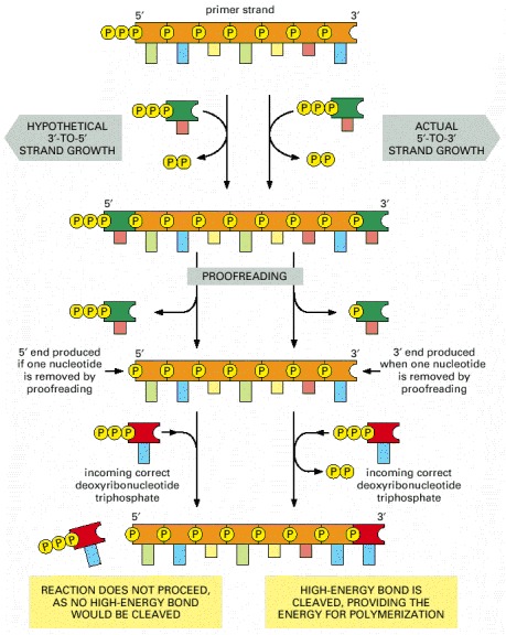
Figure 5-11An explanation for the 5′-to-3′ direction of DNA chain growth

Growth in the 5′-to-3′ direction, shown on the right, allows the chain to continue to be elongated when a mistake in polymerization has been removed by exonucleolytic proofreading (see Figure 5-9). In contrast, exonucleolytic proofreading in the hypothetical 3′-to-5′ polymerization scheme, shown on the left, would block further chain elongation. For convenience, only the primer strand of the   is shown.
Cover of Molecular Biology of the Cell
Molecular Biology of the Cell. 4th edition.
Alberts B, Johnson A, Lewis J, et al.
New York: Garland Science; 2002.
Copyright © 2002, Bruce Alberts, Alexander Johnson, Julian Lewis, Martin Raff, Keith Roberts, and Peter Walter; Copyright © 1983, 1989, 1994, Bruce Alberts, Dennis Bray, Julian Lewis, Martin Raff, Keith Roberts, and James D. Watson .
NCBI Bookshelf. A service of the National Library of Medicine, National Institutes of Health.

Thus proofreading requires unidirectional strand copying,  which in turn explains why replication is done using an otherwise complex and cumbersome "replication fork" machinery for the lagging strand. Of course evolving this macxhinery was an unlikely, difficult and individual-fitness lowering process (rather like sex), but it was necessary for all life as we know it to emerge billions of years ago. 


Friday, October 2, 2015

New "Nature" paper on molecular mechanism of transmitter release

I mentioned in class that transmitter release is mediated by 2 key molecules (or more exactly, types of molecule), the SNARES and synaptotagmin. SNARES are protein that bring the vesicle membrane and the presynaptic plasma membrane together at the active zone (where release occurs). The new paper ( Nature, 52562–67, 03 September 2015) describes an X-ray crystalographic study of the interface between the calcium-detecting molecule synaptotagmin (on the vesicle membrane) and the SNARES, which are anchored in both vesicle and plasma membranes, and come together in the narrow space between them to form a "helix bundle". When calcium binds to the synaptotagmin it, via its interface with the SNARES, triggers a contraction of the helix bundle pulling the 2 membranes together and then dragging them so they actually fuse. Here's the key diagram from the paper (which the Nature robot might censor, but you can also look at online at the University library); look particularly at parts c,d and e.


Of course one always knew, since Katz's seminal discovery of the role of calcium in transmitter release,  that the underlying molecular machinery would eventually be mapped out, but it's still very satisfying to see all the details falling into place. Sudhof, one of the authors of the new paper, had already received the 2013 Nobel Prize for his work (with Rothman and Scheckman) for his work on the molecular mechanism of exocytosis.